
|
||||||
|---|---|---|---|---|---|---|
| Echocardiography 5 minutes before starting
|
||||||
Examples of pathological findings |
||||||
|
—Echocardiographic examinations |
—Cardiac function and PA pressure |
—Examples of pathological |
||||
| echo|case | ||||||
echo|case is an echocardiographic "showcase", intended to present pregnant, visual
impressive echocardiographic findings with high didactical value, seen from the point of view and experience of different
echocardiographists.
This is not a section for case reports, namely, it is not destined for unusual echocardiographic cases or cases of
excepcional occurrence. |
||||||
|
Mobile left atrial mass |
||||||
|
Left:
Bidimensional mode, para- sternal long axis projection. The left ventricle is hypertrophic, with impaired contractility.
|
|||||

|
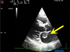
Left:
apical four-chamber view, here inverted following a Mayo Clinic tradition. The mobile mass
in the left atrium causes a partial mitral valve obstruction during diastole.
|
|||||
A differential diagnosis can be proposed: mobile thrombus in the left atrium. However, sinus rhythm make this option
more distant. The diagnosis of atrial myxoma was histologically confirmed after surgery.
|
||||||
©
Derliz Mereles |
||||||
|
echobasics | free echocardiography tutorial online since 2004 |
||||||
 ORC
ORC