
|
||||||
|---|---|---|---|---|---|---|
| Echocardiography 5 minutes before starting
|
||||||
Echocardiographic examinations |
||||||
|
—Echocardiographic examinations |
—Cardiac function and PA pressure |
—Examples of pathological |
||||
| TEE - views | ||||||
|
Guidelines and Standards Guidelines for Performing a Comprehensive Transesophageal Echocardiographic Examination, 2013 Guidelines for Performing a Comprehensive Transesophageal Echocardiographic Examination in Children and All Patients with Congenital Heart Disease, 2019 Guidelines for the Use of Transesophageal Echocardiography to Assist with Surgical Decision-Making in the Operating Room, 2020
|
||||||
|
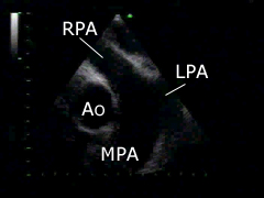
Upper position - view of the great vessels, 0°: the ascending aorta (Ao), the main pulmonary artery (MPA) and the right pulmo- nary artery (RPA) can be displayed. |
|||||
|
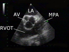
Upper position - view of the aortic valve and the pulmona- ry artery, 40-50°: the aortic valve (AV), the main pulmonary artery (MPA), the right ventricular outflow tract (RVOT) and the left atrium (LA) can be displayed. |
|||||
|
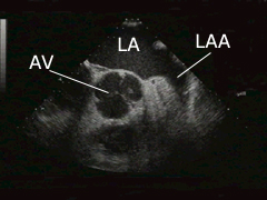
Upper position - view of the aortic valve, 40-50°: the aortic valve (AV), the left atrial appendage (LAA) and the left atrium (LA) can be displayed. |
|||||
|
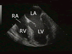
Mid-position views, 0°: the left (LV) and the right ventricle (RV), as well as the left (LA) and right atrium (RA) can be displayed. This view is similar to the four-chamber view in TTE. It is possible to get a two-chamber view (60°) and three-chamber view (120°) from this position. |
|||||
|
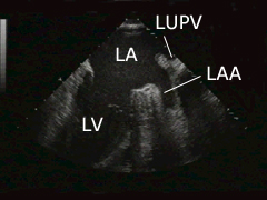
Mid-position views, 90° left: the left ventricle (LV), the left atrium (LA), the left atrial appendage (LAA) and the left upper pulmonary vein (LUPV) can be displayed. |
|||||

|
Mid-position views, 90° right: the left (LA) and right atrium (RA), as well as the right atrial appendage (RAA), the superior vena cava (VCS), the right pulmonary artery (RPA), the interatrial septum and the inferior vena cava (VCI) can be displayed. |
|||||
|
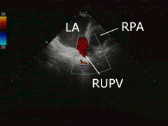
Mid-position views, 90° further right: the left atrium (LA), the right upper pulmonary vein (RUPV) and the right pulmonary artery (RPA) can be displayed. |
|||||
|
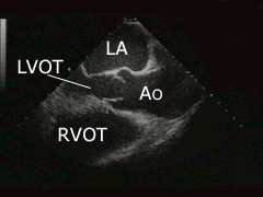
Mid-position views, 120°: the ascending aorta (Ao), the left atrium (LA), the left ventricular outflow tract (LVOT) and part of the right ventricular outflow tract (RVOT) can be displayed. |
|||||
|
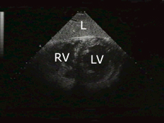
Transgastric view - short axis, 0°: the left (LV) and the right ventricle (RV), as well as the liver can be displayed. |
|||||
|
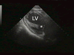
Transgastric view - long axis, 90°: the left ventricle (LV) and the mitral valve apparatus (*) can be displayed. |
|||||
|
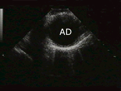
Dorsal view - during pullback, 0°: the descending aorta (AD) can be displayed. |
|||||
|
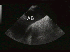
Exit view, 0°: last view before TEE probe pullback is completed. At the upper part of the ascending aorta turn the probe to the right, to display the aortic arch (AB) completely. |
|||||
©
Derliz Mereles |
||||||
|
echobasics | free echocardiography tutorial online since 2004 |
||||||
 ORC
ORC