Assessment and description of left ventricular function comprises usually its systolic or diastolic, global or
regional aspects. Myocardial function during the whole cardiac cycle is more complex, due to myocardial
architecture. Radial left ventricular function predominates certainly, but longitudinal and torsional
function also play a role. Global strain (e.g. 2D-strain), as well as other parameters, can give an
insight in the longitudinal left ventricular function. Radial LV function can be assessed with
the methods presented below.
— Calculation of left ventricular ejection fraction, LV-EF
— Formula: [(EDV - ESV) / EDV] x 100 = EF (%)
— Assessment of LV volumina with the method of discs (modified Simpson's rule, biplane)
Regional wall motion assessment
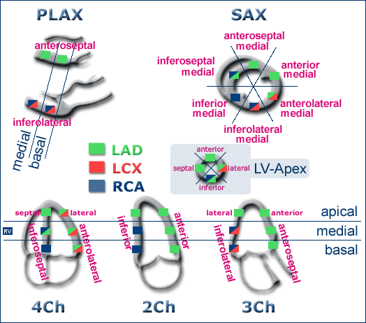
17-segment model: left ventricular wall segments
There are several models to depict left ventricular wall segments, and correspondingly, some
confusion. The 16-segment model, suggested by the American Society of Echocardiography in 1989
has proven its practicability in clinical work.
Three-chamber view, used regularly in echocardiography
examinations in Europe since decades introduced two more apical segments: anteroseptal apical and
inferolateral apical (18-segment model). In American models, apical segments remained only 4: apical anterior, apical lateral,
apical inferior and apical septal.
The typical distribution of coronary perfusion and the new 17-segment model from the ASE shown here. The 17 segment represents the so call
"apical cap". Segmental denomination has change since 2005: there are no more posterior segments, also no pure septal or lateral segments,
but anterior and inferior segments (anteroseptal, anterior and anterolateral, as well as
inferoseptal, inferior and inferolateral).
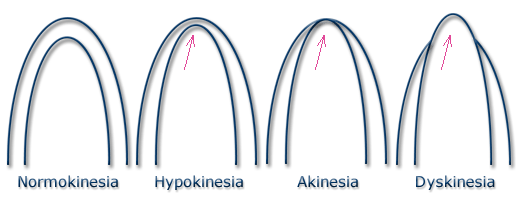
Left: normokinesia of all wall segments in four-chamber
view. Notice the slight lesser movement of septal compared to lateral segments. This is a physiological
phenomenon.
Right: lateral hypokinesia. A light increase of wall thinkness during systole
can still be seen. Notice the clear septal hyperdynamia as a compensatory reaction.
Left:
inferior basal akynesia, inferior medial hypokinesia in the two-chamber view. Notice the absence of
myocardial thickening in the akinetic segment.
Right: akinesia of the LV apex. Notice die excentric movement of the
corresponding LV segments during the systole.
Left: mild impairment of the sys- tolic left ventricular
function with hypokinesia inferoseptal.
Right: akinesia anterolateral and hypokinesia inferoseptal.
Left:
dyskinesia inferobasal with formation of an aneurysm.
Right: dilated cardiomyopathy with severe impairment of the systolic
left ventricular function.
Left: 3D volumetry of the left ven- tricle.
Offline reconstruc- tion in a case with normal LV function. 3D EF is here 73%.
Right: 3D EF of 38% here in a case with anterior wall infarction with formation of an aneurysm.
These examples were friendly provided by Dr. med. Sebastian Buss.

 ORC
ORC